A pivotal role to play
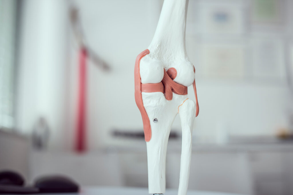
As the largest joint in the human body, the knee forms a movable connection between the thigh bone and the tibia. It connects the thigh, kneecap and tibia and is stabilised by a complex set of ligaments. As a rotational hinge joint, the knee can be flexed and rotated. The bending is composed of rolling and sliding motions. Rotation in a bent state is possible. Sliding, rolling and turning motions are enabled. During its lifetime, it may bend and stretch millions of times.
The C-shaped inner meniscus and the almost circular outer meniscus are considered to be “compensating shock absorbers.” They compensate for the inequality between the joint surfaces of the upper and lower leg and perform a certain shock absorbing function due to their soft cartilage.
A complex set of ligaments serves to stabilise the knee joint. The lateral ligaments (inner and outer ligaments) stabilise and prevent lateral bending. The cruciate ligaments are located in the joint cavity and form the shape of an X when viewed from the front. In addition, there are some ligaments that anchor the menisci, stabilise the kneecap and reinforce the joint capsule.
Range of treatments: The knee
- Meniscal injuries
- Ligament injuries
- Cartilage injuries
- Knee instability
- Patella instability
- Fracture
- Tendon injuries around the knee
- Arthrosis
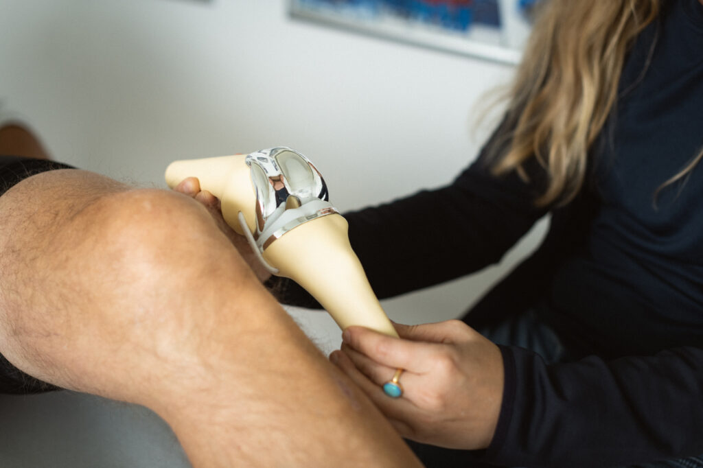
- Implantation of patial and total knee replacement
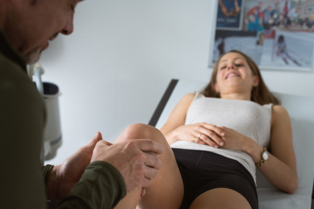
Special pathologies affecting the knee joint
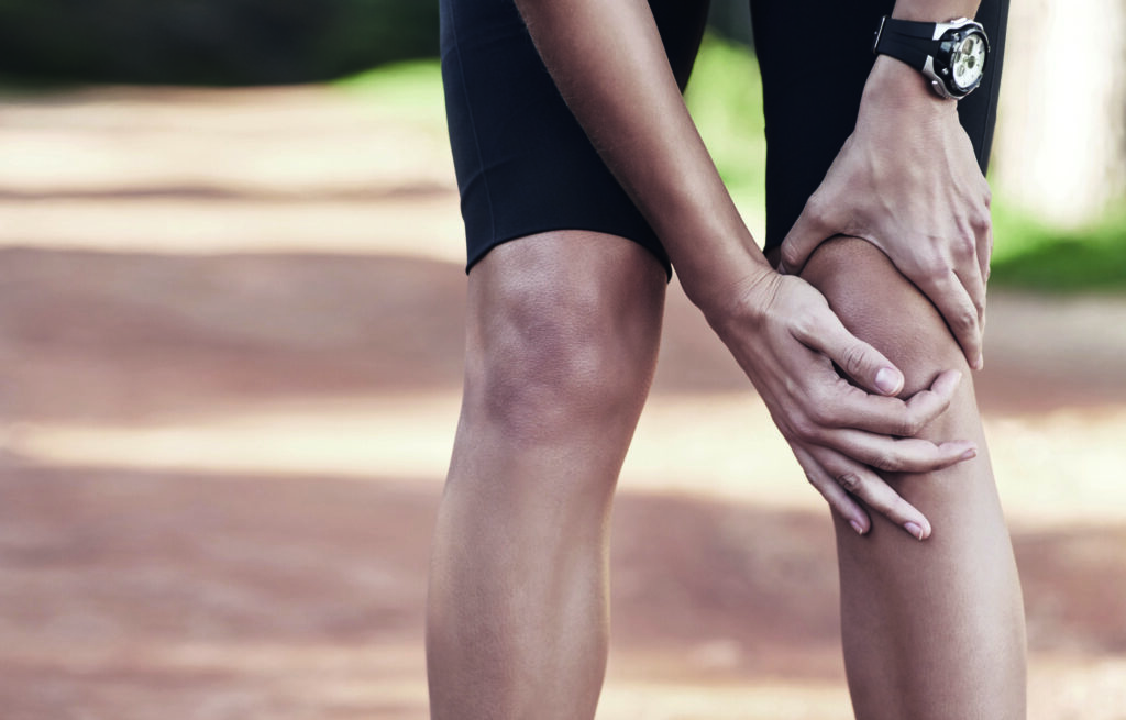
Patellar tendinitis — Jumper’s knee
Causes
The patellar tendon transmits the entire force of the largest thigh muscle to the lower leg via the kneecap. Stresses and strains inevitably lead to minute injuries (microtraumas), which normally heal without problems. However, overloading or incorrect strain may result in scarring, the excessive formation of blood vessels and swelling. If the root causes of patellar tendinitis are not treated, they can lead to chronic discomfort or even a rupture of the tendon.
Typical symptoms
Sharp pain during and after putting weight on the knee or tenderness along the tendon or the areas around the tendon are characteristic. Visual swelling is often present at the same time.
Therapy options
- Active strengthening
Targeted strength training is an indispensable way to strengthen overstrained muscles and to prevent pain from reoccurring. - Stretching
Regularly stretching the affected muscles minimises tension and positively affects the healing process. - Knee alignment training
This results in a significant load reduction in the knee joint area. - Targeted massages
Self-massage can eliminate muscle stiffness and improve blood circulation. - Anti-inflammatory medication
Either as an ointment, gel or spray on the skin, or taken in pill form, anti-inflammatory drugs contribute greatly to improving symptoms. - Shockwave therapy
Concentrated pressure waves promote local blood circulation, have a beneficial effect on inflammatory processes, and help to relax the muscles. - AlterG
Running on an “anti-gravity treadmill” has a positive effect on healing. - Injections
For long-lasting pain, the injection of activated autologous blood (ACP) is often a consideration. - Surgery
During surgical procedures, open or arthroscopic, the patellar apex is rounded off. Dead tissue can be removed at the same time.
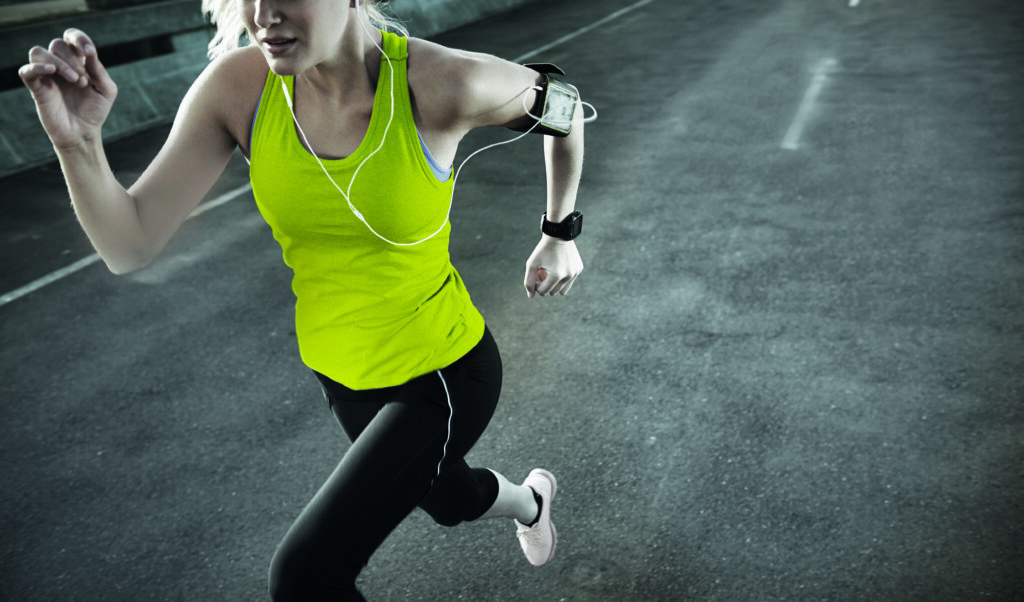
Iliotibial band syndrome — Runner’s knee
Causes
When the knee joint is flexed, a broad tendon plate (iliotibial tract) slides over the outside of the thigh bone just above the joint. Monotonous movements (running, cycling) can lead to overuse injuries in this area. Triggering factors can be leg axis malpositions, unfavorable surfaces, poor footwear and training errors. Often such injuries can be attributed to weakness or a shortening of the gluteus medius muscle. The aim of the treatment is to correct the muscle imbalance and stabilise the leg axis in order to achieve a normal sequence of motion.
Typical symptoms
Sudden, severe, stabbing pain on the outside of the knee joint that occurs after 15-30 minutes but subsides relatively quickly.
Therapy options
- Active strengthening
Targeted strength training is an indispensable way to strengthen overstrained muscles and to prevent pain from reoccurring. - Stretching
Regularly stretching the affected muscles minimises tension and positively affects the healing process. - Knee alignment training
This results in a significant load reduction in the knee joint area. - Targeted massages
Self-massage can eliminate muscle stiffness and improve blood circulation. Use a Blackroll or similar to release fascia adhesions and reduce tension. - Anti-inflammatory medication
Either as an ointment, gel or spray on the skin, or taken in pill form, anti-inflammatory drugs contribute greatly to improving symptoms. - Shockwave therapy
Concentrated pressure waves promote local blood circulation, have a beneficial effect on inflammatory processes, and help to relax the muscles. - Injections
If the pain persists for a long time, an injection of corticosteroid may be considered. Unlike other tendons, there is no risk of rupture. - Surgery
During surgical procedures, open or arthroscopic, the iliotibial tract is operated upon, thereby reducing tension. Dead tissue can be removed at the same time.